
Visual health

Retina

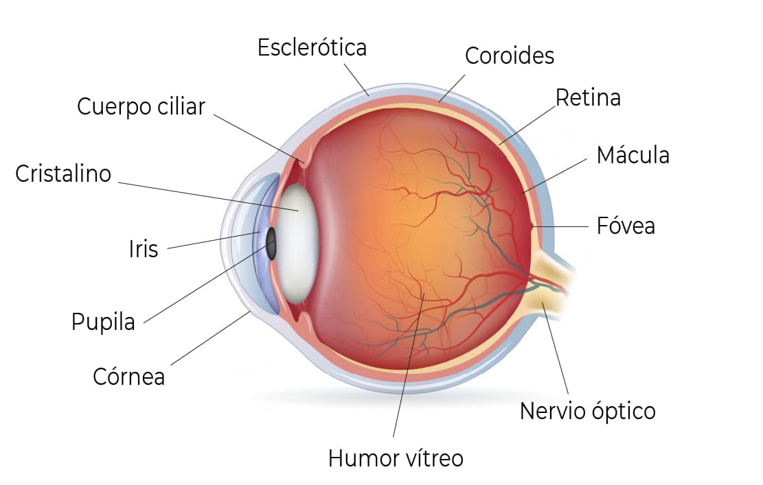
The retina is the inner membrane of the eye that receives light impressions and transmits them to the brain; it covers the choroid up to the iris and is made up mainly of expansions of the optic nerve.

Anatomy of the eye and its parts

Retinal detachment

Retinal detachment (RD) is a process in which the sensory retina separates from the retinal pigment epithelium due to the presence of subretinal fluid. It is a medical emergency requiring surgical treatment, with a guarded visual prognosis.

An annual dilated eye exam is advisable to detect retinal lesions considered predisposing factors for retinal detachment. Lesions caught early require close follow-up and, eventually, laser photocoagulation treatment.

Floaters

The perception of "floaters" — small moving spots, threads or other shapes drifting across your vision — is one of the most common reasons for an eye exam, and is medically known as vitreous floaters (miodesopsias).

These floaters characteristically move along with your eye movements. In most cases they are related to physiological changes in the vitreous humor and are harmless, but in some cases they can be associated with serious eye conditions that require specific treatment.

The main risk factors are advancing age and myopia.

Treatment

Floaters usually don't require treatment. In cases linked to an evolving underlying condition, follow-up and treatment will be indicated as needed.

Diabetic retinopathy

Diabetic retinopathy is a common complication in diabetic patients. High blood glucose levels damage the retina's blood vessels, causing fluid leaks (edema) or vessel closure (ischemia), which harms the retina. In advanced stages of the disease, abnormal new vessels form and eventually cause retinal detachment — a stage known as proliferative retinopathy.

Treatment

Diabetes is a silent, multisystem disease. Teamwork with a physician and an endocrinologist is essential, since good control prevents or delays complications. Retinal damage at any stage reflects poor disease control.

Age-related macular degeneration

Age-related macular degeneration is the aging or deterioration of the retina's central visual area — the macula — and is directly linked to age. It is defined by choroidal neovascularization and/or geographic atrophy (a sharply demarcated area of the pigment epithelium) along with drusen (yellowish spots in the neuroretina).

This condition is more common after age 55, more prevalent in women, and more likely in smokers, people with hypertension, or those with a family history of macular degeneration.

Macular edema

The retina has two regions: the central retina and the peripheral retina. The central retina has a small depression (about 1mm x 1.5mm) called the macula, made up entirely of highly specialized cells called cones, which allow detailed and color vision. The macula lets us read, write and see the fine details of objects. Any disease affecting this area is called maculopathy. The accumulation of fluid in this central retinal area is known as macular edema.

An eye exam is recommended for anyone with a family history.

Retinitis pigmentosa

Retinitis pigmentosa is a genetic retinal dystrophy. It has two forms of inheritance: recessive, the more severe form, and dominant, which is benign. Many of the responsible genes can be identified through genetic mapping.

It typically appears between ages 25 and 40 and is usually noticed as difficulty with night vision. An eye exam is recommended for anyone with a family history.

Treatment

There have been advances in genetic studies and, in the future, in mapping all of the genes involved. Rheological treatments are currently used to slow damage and progression. Another option under study is stem cell transplantation in the retina, though there is still a long way to go.

Want to get in touch with us?

You can call or message us on WhatsApp at +549 11-5823-7463 (WhatsApp and calls), or call +549 11-5823-2375 (calls only).